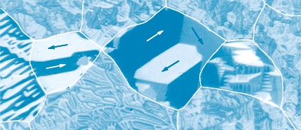
|
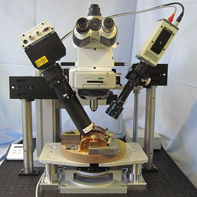
The combined system is a highly flexile setup that combines two microscope versions:
(i) a low-resolution version and
(ii) a high-resolution optical microscope.
Both versions are implemented in the same setup and can be used optionally.
Features (excerpts):
|
|
Polarization Microscopes
|
Two optional versions of wide-field Kerr microscopes are implemented in the setup:
Version 1: low-resolution optical microscope
Allows to obtain an overview of the domain pattern (or magnetization loop) of larger samples with a spot size from approx. 30 by 30 mm down to 10 by 10 mm (zoom optics) Separated illumination and reflection paths Rotatable polarizer, analyzer and compensator module
Version 2: high-resolution optical microscope
Allows observation down to resolution limit of optical microscopy (300 nm) Wide-field optical polarization microscope (Zeiss optics) Microscope head can be vertically shifted by approx. 150 mm to allow optional use of Version 1-microscope Objective lenses with magnifications from 5x to 100x (oil immersion). Optical zoom lenses 1x, 2.5x, and 4x for increasing the magnification of the microscope image (not the resolution!) Computer controlled choice of Kerr sensitivity. Depending on the configuration, the following Kerr sensitivities can be chosen:
(i) longitudinal sensitivity with superimposed polar sensitivity,
(ii) transverse sensitivity with superimposed polar sensitivity,
(iii) pure polar sensitivity,
(iv) simultaneous display of longitudinal and transverse contrast (with polar contrast superimposed),
(v) simultaneous display of pure longitudinal and pure transverse contrast,
(vi) simultaneous display of pure longitudinal (or pure transverse) contrast and pure polar contrast
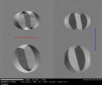
Polarizer for increased Kerr-efficiency, rotatable by approx. 100° Rotatable analyzer and compensator module Sample stage for sample sizes up to 35 mm, manual x-y sample shift up to ± 14 mm, manual sample rotation by 360° Computer-controlled compensation of parasitic Faraday effect in objective lens
|
|
Light Source
|
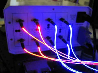
Overview microscope: highly stable LED light source (white light - monochromatic light on request) High-resolution microscope: specially developed Kerr-LED lamp, based on eight light-emitting diodes the light of which is guided to the microscope by glass fibers. The ends of the fibers are arranged in a specific array-way and are imaged to the diffraction plane of the microscope.
Various Kerr-sensitivity options (longitudinal, transverse, and polar Kerr sensitivities) can be chosen by activating different LEDs of the array. The selection is made conveniently by computer control.
The Kerr-LED lamp can be obtained in a monochromatic (white light) or dichromatic (blue and red light) version. In this latter high-end version, images of different color and Kerr sensitivity are generated at the same time and separated by a color-sensitive image splitting device between microscope and camera. Both images are then displayed simultaneously within the same frame on the screen.
|
|
Sensitive low noise digital (cooled) CCD camera
|
|
|
Rotatable Electromagnet ("Standard" Magnet)
|
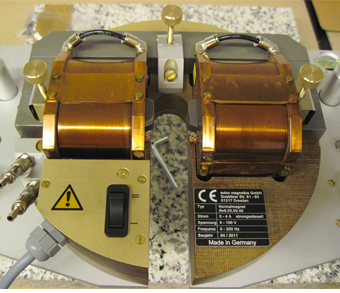
Manually rotatble by approx. 360° Supported on a magnet holder with adjustable height, support independent from sample holder Water-cooled In-plane field range from 10-4 T up to 1.40 T
|
|
Measurement Computer and Software
|
Configurated workstation NI LabView based measurement software "KerrLab" Camera control Magnetic field control (DC, AC, demagnetization etc) Image processing capabilities for high contrast magnetic domain observation (real-time difference imaging technique) Recording of surface hysteresis loops (MOKE magnetometry) with simultaneous domain movement observation
|
|


